August 2025 TPJ Editor choice: The making of a leaf tip: how cell division angles define shape
Highlighted publication:
Biregionally differentiated growth generates sharp apex and concave joints in leaves
https://doi.org/10.1111/tpj.70310
The making of a leaf tip: how cell division angles define shape
Leaves come in an extraordinary range of shapes: they can be simple or dissected, can have smooth or toothed margins, and can display a variety of lobes, points and indentations. These shape variations have important functional consequences, affecting light capture, water loss, wind resistance, and even herbivore interactions. Leaves originate as small primordia on the flanks of the shoot apical meristem, from which they grow and expand until they reach their mature form. The rate, duration, and spatial patterning of cell division and expansion are key determinants of leaf size and shape, but how these processes are coordinated is not well understood.
Wang and colleagues took on the challenge of dissecting leaf shape formation in Triadica sebifera – a member of the spurge family and an oil-producing species of economic significance. Its leaves are distinctly shaped, featuring a rounded base and a sharply tapering tip, linked by concave joint regions (Fig. 1a). This shape can be quantified by its curvature, i.e., how much the contour deviates from a straight line (Fig. 1b): the base shows moderately positive curvature (convex, rounded), the joint regions display negative curvature (concave), and the tip reaches a maximum curvature with a sharp point. Until now, it has remained unknown how this distinctive apex forms.
Wang et al. initially modelled leaf growth using contour growth mapping, moving contour points of the leaf outline outward at defined speeds. When growth was isotropic (equal in all directions), the model produced a circular leaf. Vertical anisotropic growth yielded an elliptical form. However, combining isotropic growth at the base with vertical anisotropic growth at the tip generated a shape resembling T. sebifera leaves, indicating that distinct regional growth patterns are required.
To assess whether this biregional growth is reflected at the cellular level, Wang and colleagues analysed cell shapes and division patterns during leaf development. Interestingly, the sharp apex was already present in very young primordia, despite apical cells lacking elongation, and no elongation was observed after this stage. This indicated that cell elongation was not responsible for tip formation. Instead, the orientation of cell division emerged as a key factor. Division angles were vertically aligned (tip-to-base) in the apex, leading to horizontally oriented cells, whereas angles at the base changed from vertical to random orientation as the primordium matured.
These findings prompted Wang et al. to test whether a biregional pattern of cell division angles alone could generate the observed leaf shape. They turned to a more detailed vertex model, which simulates cellular growth by representing cells as polygons, and cell divisions as the splitting of these shapes. Random division angles yielded a circular form, while uniformly vertical divisions created a rod-like shape. However, fixing the division angle to be vertical only in the apex region produced a form closely resembling T. sebifera leaves (Fig. 1c).
Spatial control of division angles thus appears sufficient to explain T. sebifera’s leaf shape. Yet Wang’s previous observations showed that division angles in the base region change over time and that an “arrest front” forms, progressively restricting division to the basal region of the leaf. This suggested a temporal component to leaf shape control. The authors therefore incorporated this idea into their model, adding dynamic changes in division orientation and an advancing arrest front. Remarkably, this version of the model was also able to reproduce the characteristic leaf shape, even without predefined regional differences in the division angle (Fig. 1d).
In conclusion, Wang et al. show that both spatial and temporal regulation of division angles can generate leaves with sharply pointed tips. The authors view these not as competing hypotheses, but as complementary aspects of a unified developmental process, where spatial patterns represent the outcome of shifting division angles and division activity over time.
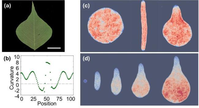
Figure 1. Spatial and temporal changes in cell division angle can generate the leaf shape of Triadica sebifera.
(a, b) Representative image of a T. sebifera leaf (a) and the corresponding curvature plot of its outline (b).
(c) Examples of organs generated from numerical simulations of different cell division angles: random angles (left), exclusively vertical angles (middle) and a combination of vertical angles in the apical and random angles in the basal region (right). Colour indicates division numbers (red – high; blue – low).
(d) Time series of simulations combining an advancing arrest front and temporally regulated division angles. Initially, all cells divide in a vertical direction, while later, only basal cells divide with random division angles. Colour indicates division numbers (red – high; blue – low).
Figure modified from Wang et al. (2025).